
ENIGMA-Parkinson’s Disease (ENIGMA-PD) is an upcoming disease working group led by Dr. Boris Gutman of USC. The ENIGMA-PD project will consist of four major studies, including:
- A meta-analysis of volumetric changes in Parkinsonian versus healthy brains, using previously developed FreeSurfer protocols
- A meta-analysis of white matter changes, based on TBSS and our customized ENIGMA template and atlas
- A shape meta-analysis, based on the shape analysis pipelines developed by Dr. Gutman and colleagues
- Multi-site voxel-based meta-analysis, which is currently being tested by Dr. Neda Jahanshad.
ENIGMA-PD is presently comprised of two major datasets from (i) the Moscow Research Center for Neurology (PI – Sergei Illarioshkin) and (ii) the Parkinson’s Progressive Markers Initiative (PPMI). Results from our preliminary analysis the PPMI dataset are provided below.
If you are interested in joining ENIGMA-PD, please contact Boris Gutman at bgutman@gmail.com.
Striatal and Thalamic Shape Alterations due to Parkinson’s Disease
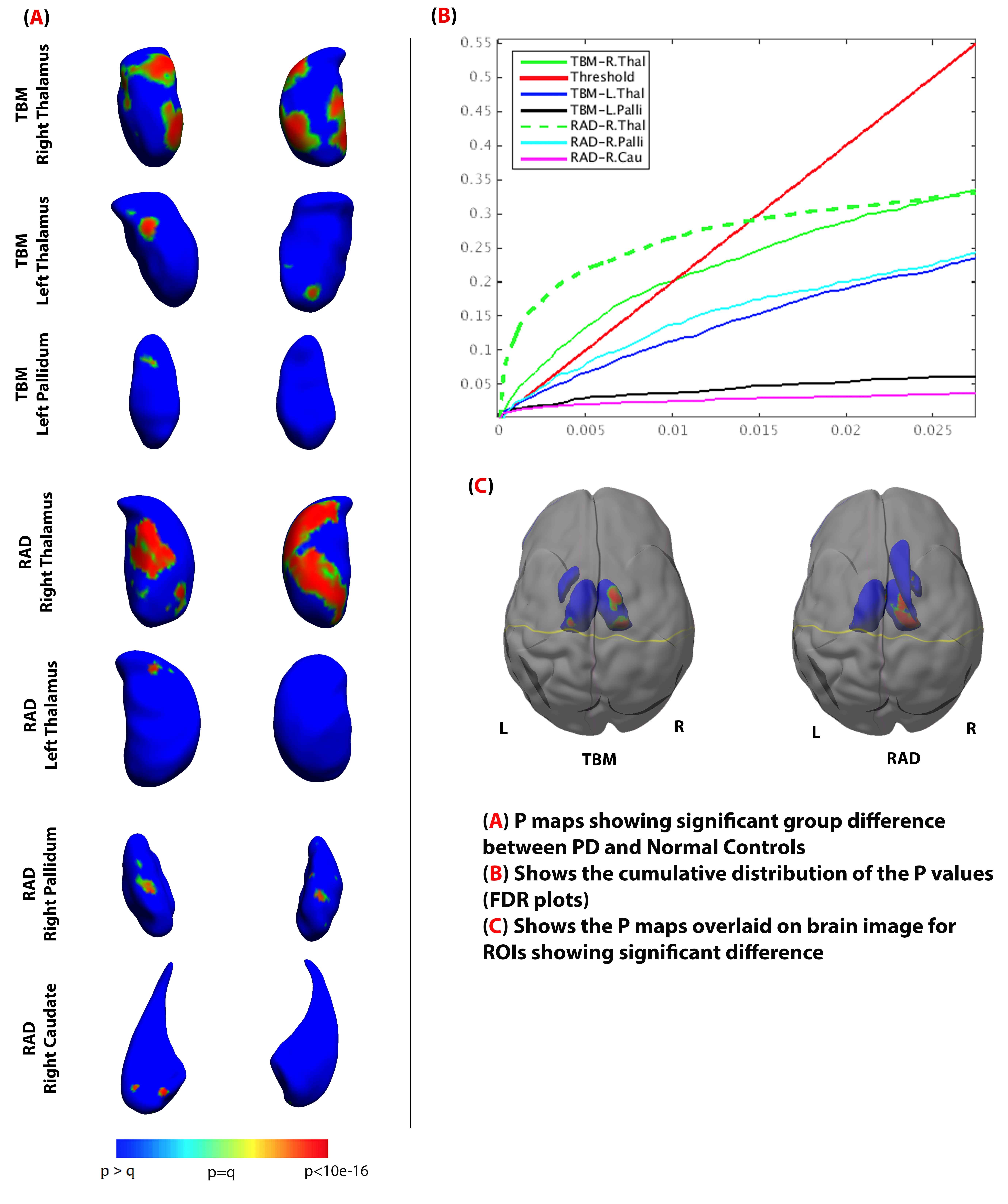
Anjanibhargavi Ragothaman, Christopher Ching, Zvart Abaryan, Adam Mezher, Paul Thompson, Boris GutmanParkinson’s disease (PD) is a progressive neurological motor related disorder, characterized by trouble initiating movement, resting tremors, slowness of movement and muscular stiffness [1]. Volumetric analysis of the brain images tends not to reveal subtle changes in structure for patients with PD. Alternatively, shape analysis of subcortical region boundaries may reveal more subtle, localized effects. In this study, we used a medial demons surface registration and shape analysis of 7 subcortical regions: amygdala, nucleus accumbens, putamen, caudate, globus pallidus, hippocampus, and thalamus. Following shape registration, two surface measures – thickness and dilation ratio (Jacobian Determinant) – were used to quantify local shape differences [2].
The data were obtained from the Parkinson’s Progressive Markers Initiative (PPMI) (116M:68F healthy controls and 264M:142F PD subjects. Subcortical parcellations were obtained using Freesurfer 5.3. Mass-univariate test for group difference between controls and PD subjects was performed, controlling for age, gender, ICV and handedness. Shape signatures of the following regions were significantly different after False Discovery Rate (FDR) correction for multiple comparisons (critical q in parentheses, higher implies greater effect): right and left thalamus (q=0.0102, q=0.001), left pallidum (q=3.39e-04) for dilation ratio measures. Right and left thalamus (q=0.00144, q=1.69e-04), right pallidum (q=0.0012) and right caudate (q=4.76e-04) showed significant difference for radial thickness measures. P-maps localizing group differences are shown in Figure 1, and are consistent with previous findings in PPMI [3]. This study suggests that subcortical shape changes can be used as biomarkers for early detection of the disease.
- Rivlin-Etzion M, Elias S, Heimer G, Bergman H: Computational physiology of the basal ganglia in Parkinson’s disease. Prog Brain Res 2010, 183:259-273.
- Gutman BA, Jahanshad N, Ching CRK, Wang Y, Kochunov PV, Nichols TE, Thompson PM: Medial Demons Registration Localizes the Degree of Genetic Influence over Subcortical Shape Variability: An N= 1480 Meta-Analysis. International Symposium on Biomedical Imaging (ISBI 2015) 2015.
- Garg A, Appel-Cresswell S, Popuri K, McKeown MJ, Beg MF: Morphological alterations in the caudate, putamen, pallidum, and thalamus in Parkinson’s disease. Frontiers in Neuroscience 2015, 9:101.